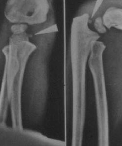
An avulsion of the medial epicondyle, also called a medial epicondyle fracture, commonly occurs in the pediatric population between the ages of 9-14 years old. This accounts for about 20% of all pediatric elbow fractures. This fracture has become more common due to the increase in the athletic demands of the pediatric population. The injury is referred to as an avulsion injury because it occurs when there is excess valgus stress with contraction of the flexor-pronator mass. The mechanism of the injury also makes it very common to occur with elbow dislocations as well. While most of these dislocations will spontaneously reduce before making it to a hospital, the medial epicondyle bony fragment can get incarcerated in the joint during the reduction. Figure 1 shows an example of an incarcerated medial epicondyle in the elbow joint after associated elbow dislocation.

Image Courtesy - https://www.orthobullets.com/pediatrics/4008/medial-epicondylar-fractures--pediatric
Figure 1
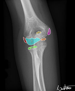
The elbow has six ossification centers, and the medial epicondyle is the last one to fuse which makes this injury common in adolescents when the ossification center is weakest. This ossification center does not contribute to longitudinal growth so injuries to the medial epicondyle do not cause a significant length discrepancy between the two arms; however, it can cause a painful nonunion if it does not heal, especially in throwing athletes. Figure 2 shows the 6 ossification centers in the elbow with “I” indicating the medial epicondyle. The blood supply comes from branches of the inferior ulnar collateral artery and branches of the superior and inferior ulnar collateral artery.

Image Courtesy - https://radiopaedia.org/articles/ossification-centres-of-the-elbow?lang=us
Figure 2
The common flexor-pronator wad attaches to the medial epicondyle, which is responsible for pulling the medial epicondyle off during the valgus force. The muscles included in this wad are the pronator teres, flexor carpi radialis, palmaris longus, flexor digitorum superficialis, and the flexor carpi ulnaris. These patients will present with valgus instability, ecchymosis, generalized swelling in associated elbow dislocations, and occasionally ulnar nerve dysfunction.
Treatment can include both operative and nonoperative measures depending on the nature of the fracture. The fracture can be treated with immobilization in a long arm cast flexed at 90 degrees for 1-3 weeks if the fracture is less than 5mm displaced. Operative management is indicated when there is displacement of the fracture greater than 5 mm, entrapment of the fracture fragment in the joint space, extension of the fracture to the articular surface, or an open fracture. Displacement can be very hard to determine off regular radiographs and so sometimes a CT scan is obtained of the elbow to determine appropriate management.

Image Courtesy - https://www.orthobullets.com/pediatrics/4008/medial-epicondylar-fractures--pediatric
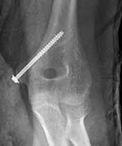
Figure 3
Figure 3 shows the most common mechanism of fixation. An incision is made directly over the medial epicondyle and care is taken to identify and protect the ulnar nerve. The fracture is reduced and then a k-wire is placed to hold the reduction. A cannulated screw is then placed over the k-wire and then the k-wire is removed. There is very little soft tissue around the elbow which can lead to prominent hardware. This hardware can become bothersome for many patients and may require removal in the months following surgery once the fracture is healed.


