The articular cartilage in the knee, made up of mostly type 2 collagen, covers the ends of the femur and tibia in your joint. Cartilage lesions in the knee comprise a spectrum of entities from single localized defects to broader advanced degenerative disease also known as osteoarthritis. More localized and contained cartilage lesions in the weight bearing surface of the knee can be treated with procedures referred to as cartilage restoration procedures. What specifically is done in these procedures depends on the size and depth of the lesion.
What causes cartilage defects?
Either acute trauma or chronic repetitive overload can cause cartilage defects. Oftentimes, the mechanism of injury is unknown for the specific osteochondral lesion. Osteochondral lesions can be associated with acute ligamentous injuries such as an ACL tear, or an undiagnosed osteochondral defect that occurred as a child and presents itself later in life. Usually, a force is applied to your joint that causes the femur and tibia to come in contact with each other at such a force that it damages the articular cartilage.
Can a cartilage defect heal itself?
Articular cartilage lesions have a limited ability to heal when compared to other tissues in the body such as tendons or muscle injuries. This is because cartilage is avascular and relatively acellular when compared to other tissues in the body. This limits its spontaneous healing capacity. Since cartilage does not heal itself well, over time it tends to worsen and break down further.
How do you treat cartilage lesions?
How you treat cartilage lesions depends on the size and depth of the lesion. With smaller lesions, either simple debridement or microdrilling procedures which restore blood supply to the injured area are often sufficient for the body to promote a healing response. With acute injuries, sometimes the cartilage piece can be fixed back onto the injury site. Other techniques can involve using viable cartilage allograft such as Cartimax. However, if the lesion is larger (such as 15-30 mm), an osteochondral allograft is often used to fill in that cartilage defect.
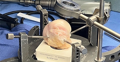
Figure 1 shows a large ostochondral defect that is prepared for transplant surgery. This type of surgery is a much larger surgery and requires a prolonged healing period. Osteochondral allografts are taken from a donor’s body and they are specifically cut to fill in the defect in the knee. This defect is usually on one of the femoral condyles. Figure 2 shows the defect filled with an osteochondral allograft. Overtime, this allograft will incorporate into the knee and help distribute force evenly across the joint.
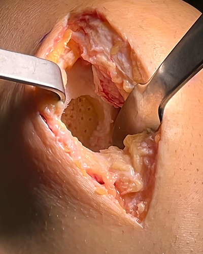
Figure 1
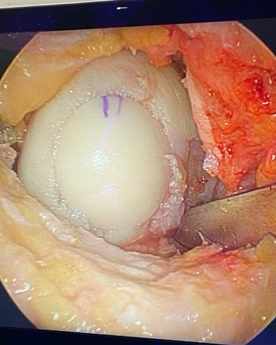
Figure 2
How long does it take to recover from a cartilage procedure?
It typically takes an active individual several months and up to a year to be able to return to full activity after a cartilage restoration procedure. For the first few weeks after surgery, the patient will be heel touch weight bearing in an extension brace with crutches. After this, the patient will be able to progress weight bearing on their leg and wean off the brace but must not undergo heavy loading activities such as jumping or running. Typically, the patient can start advancing to their sports related activities after six months.