

Anterior cruciate ligament (ACL) tears are among the most common knee injuries seen in active, cutting athletes. They occur when the ACL, one of the most important stabilizing ligaments of the knee, is overstretched or torn. Prompt diagnosis is crucial for making decisions regarding treatment. Below is a step by step guide on how Orthopaedic Surgeons diagnose these injuries.
History and Physical Exam
The diagnostic process begins with a review of the events surrounding the injury or the development of symptoms. Physicians will ask detailed questions about the event such as:
How did the injury occur? (pivot, awkward landing, or collision)
Did you hear or feel a “pop”?
Are you having swelling, instability, or difficulty bearing weight?
Symptoms of an ACL tear include knee swelling, instability (a feeling that the knee might give out), and restricted range of motion. Pain levels can vary but are usually moderate immediately following the tear.
Following the history, a detailed physical exam is performed. Physicians typically use the following tests to assess the ACL:
- Lachman Test: The patient’s knee is bent slightly to approximately 20 to 30 degrees. The examiner stabilizes the thigh and pulls the tibia forward to check for laxity, which indicates a tear.
- Anterior Drawer Test: With the knee flexed to 90 degrees, the examiner pulls the tibia forward. Increased movement compared to the uninjured knee suggests a tear.
- Pivot-Shift Test: A more specialized test that again tests for ACL tear and is done by flexing the knee while the tibia is in internal rotation. The knee will pivot as the tibia reduces with flexion. This test can be painful and difficult to perform on an awake patient and is typically done under anesthesia in the operating room or in cases with chronic ACL tears.
Imaging
While the physical exam is very important for diagnosis, the following imaging studies are often utilized to confirm history and physical exam findings:
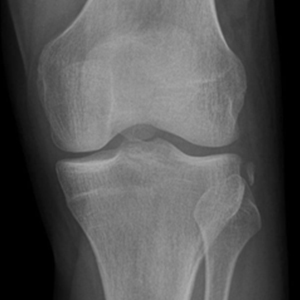
Figure 1
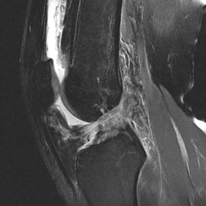
Figure 2
- X-rays: Radiographs can be very important for diagnosing ACL tears. First, they allow the physician to rule out fractures that may be leading to the patient’s symptoms. There are also signs on x-ray, including a “segond” fracture, which is a small avulsion of the capsule that is almost always associated with an ACL tear (see figure 1). One can also perform stress xrays in the setting of a multi-ligament knee injury.
- Magnetic Resonance Imaging (MRI): MRI is the gold standard with imaging for diagnosing ACL tears. MRI provides a detailed view into the soft tissues of the knee including ligaments, cartilage, and menisci. This is important because ACL tears are often associated with injuries to other structures. Figure 2 shows an ACL tear on the MRI.
In conclusion, diagnosing an ACL tear involves a combination of history, physical exam, and imaging studies. Prompt diagnosis is critical for treating patients with these injuries and getting them back to their active lives.
Please see your board certified orthopedic surgeon when seeking care for a knee injury.